Skull models
Skull models
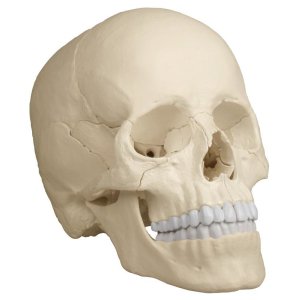
Galaxymed offers a big range of high standard skull models which are a close to real copy of human skulls for your anatomy studies. You will find a big variety of these outstanding eductorial aids.
Items 1 - 20 of 57
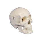
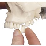
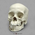
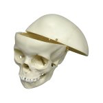
The mandible is open and shows roots, spongiosa, nerve canal and an impacted wisdom tooth. The teeth in the upper and lower jaw can be extracted and reinserted.
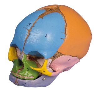
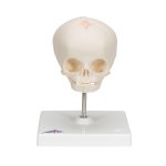
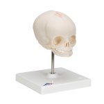
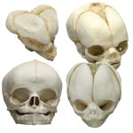
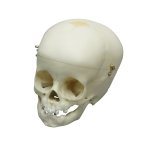
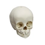
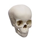
3B Scientific Smart Anatomy foetal skull model, natural cast, 30th week of pregnancy shows the characteristics of prenatal development. Foetal skull on stand.
This model gives an impressive overview over the development of the skull in utero.
Due to the very special production technology even smallest details are reproduced and the model looks and feels almost like a real human skull.
Due to the very special production technology even smallest details are reproduced and the model looks and feels almost like a real human skull.
Due to the very special production technology even smallest details are reproduced and the model looks and feels almost like a real human skull.
Due to the very special production technology even smallest details are reproduced and the model looks and feels almost like a real human skull.
Due to the very special production technology even smallest details are reproduced and the model looks and feels almost like a real human skull.
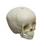
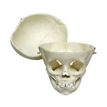
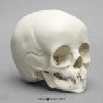
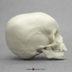
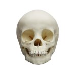
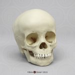
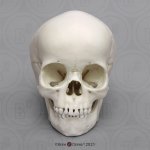
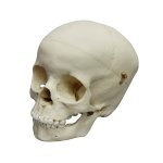
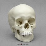
First class actual cast of a 3 year old child in extraordinary high detail. This skull also shows numerous Wormian (sutural) bones found along the lambdoid suture. The epipteric bone, which is a Wormian bone found at the anatomical landmark known as the pterion is also shown.
Due to the very special production technology even smallest details are reproduced and the model looks and feels almost like a real human skull.
Due to the very special production technology even smallest details are reproduced and the model looks and feels almost like a real human skull.
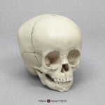
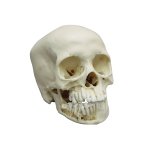
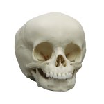
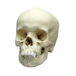
First class actual cast of a 9 year old child in extraordinary high detail. Due to the very special production technology even smallest details are reproduced and the model looks and feels almost like a real human skull.
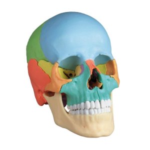
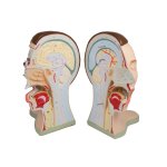
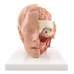
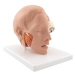
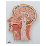
Representation of the head (differentiated in colour), medially divided.
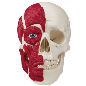
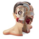
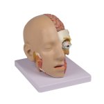
Representation of a head, medially divided. The skin and facial muscles of the right outer half are removed to show the deeper structures.
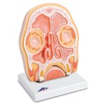
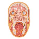
3B Scientific Smart Anatomy frontal section model of human head through the paranasal sinuses covered with mucous membrane. Signs of sinusitis on the right.
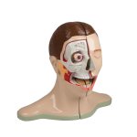
3B Scientific Smart Anatomy human head model, 6 part features a removable 4-part brain half with arteries. The eyeball with optic nerve is also removable.
The left side of the face is dissected in sagittal and horizontal section, showing many features of the skull and brain, as well as the oronasal cavity.
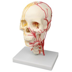
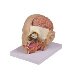
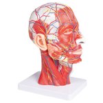
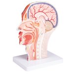
Life size model shows the right half of the human head and neck, sectioned along the sagittal plane. A superficial dissection exposes the facial muscles, the superficial blood vessels and nerve branches of the face and scalp, and the parotid and submandibular glands. A median dissection exposes the brain with its internal structure; the pharynx and upper respiratory tract; and a section of the cervical vertebrae.
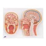
3B Scientific Smart Anatomy median and frontal section of human head. The 2 relief models show the median and frontal section of the head on baseboard.
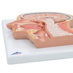
3B Scientific Smart Anatomy median section of the head. This relief model shows all relevant structures of the human head in great detail.
Items 1 - 20 of 57

