Respiratory System
Respiratory System
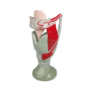
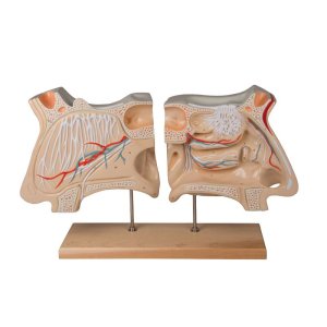
Here you find detailed close to real models or schematic display of the human respiratory system: Larynx, Lung, and Nose models.
Items 1 - 20 of 20
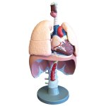
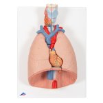
Representation of the human respiratory system in about half life size. Lungs, trachea and upper respiratory tract are shown in detail.
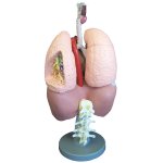
A life size model, finely colored to show all major anatomical detail. Supplied complete on stand, with key card.
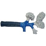
A model of the terminal bronchiole system of the lungs.
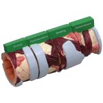
The 4 piece Bronchus model is a four stage cross-section of the bronchus demonstrating the tissue changes occurring in asthma and chronic bronchitis.
3B Scientific Smart Anatomy COPD bronchial tissue model impressively shows the changes to the bronchial tissue: normal, abnormal, secretion, thickening…
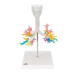
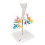
3B Scientific Smart Anatomy CT Bronchial Tree Model with Larynx is a unique way to study the anatomy of the human lungs.
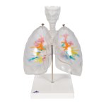
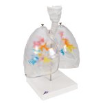
3B Scientific Smart Anatomy CT Bronchial Tree Model with Larynx & Transparent Lungs. The transparent lungs are detachable from the bronchial tree and larynx.
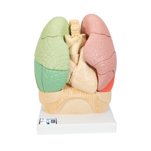
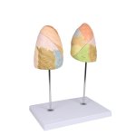
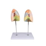
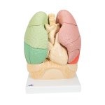
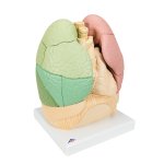
This life size model shows clearly the segmental anatomy of the left and the right lungs. The various lobes are painted with different colors to highlight their locations.
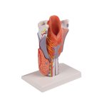
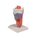
This 5-part model is medially sectioned and shows all internal structures, like the hyoid bone, cartilages, ligaments, muscles, vessels, nerves and thyroid gland.
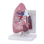
Full size lung model with normal cut-away of right side of lung shows bronchus, arteries, vein, two lymph nodes, bronchial passages and trachea bifurcation.
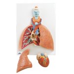
3B Scientific Smart Anatomy Lung Model with Larynx, 5 part. A great educational tool for teaching-learning the anatomy of the human lung and surrounding area.
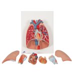
3B Scientific Smart Anatomy Human Lung Model with Larynx, 7 part is a great model of the anatomy of the lung area. The lung model with larynx is on baseboard.
3B Scientific Smart Anatomy segmented lung model shows the lungs with representation of the bronchial tree close to the heart, trachea, oesophagus and aorta.
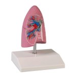
Model of a right lung in about 1/2 life size with bronchus, arteries and veins.
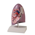
Model of a right lung in about life size with bronchus, arteries and veins.
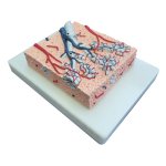
This model shows an approximately 20 times enlargement of a section through the lungs.
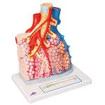
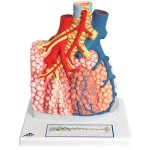
3B Scientific Smart Anatomy Model of Pulmonary Lobule with Surrounding Blood Vessels shows an external pulmonary lobe with a magnification of 130x.
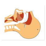
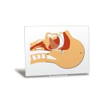
The Airway Simulation Board allows instructors to effectively demonstrate a closed and open airway.
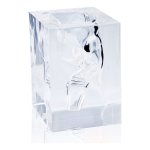
This is a transparent model that reproduces the laryngopharynx three-dimensionally, from the oropharynx to the hypopharynx. It is useful in explanations to help people understand the complex structure of the laryngopharynx, and can be used as a model when training personnel involved in swallowing therapy or when giving explanation to patients or their families
Using this transparent model, it is possible to understand at a glance and explain simply the complex structure of the nasal cavities.
Items 1 - 20 of 20


