Digestive system
Digestive System
The knowledge of the human digestive system can be easier provided with the use of anatomical models: the whole system on a schematic display from tongue to rectum or the detailed models of the single organs on its way: e.g. esophagus, insestine, stomach, kidney. liver. The models are shown in natural and with diseases.
Items 1 - 20 of 44
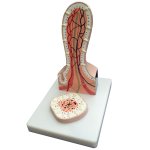
This greatly enlarged 2-part model of a villus from the small intestine shows detail from a transverse and longitudinal section.
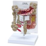
Cut-away view model with common pathologies as: adhesions, appendicitis, bacterial infection, cancer, Crohn’s disease, diverticulitis, diverticulosis, polyps…
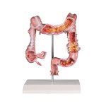
Reduced size model of a human colon showing ileum, caecum, ascending colon, transverse colon, descending colon, sigmoid colon and rectum.
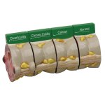
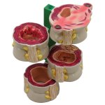
Four-piece colon model with cross-section of the human colon demonstrating both normal and various disease conditions.
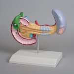
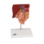
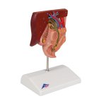
This full size model shows pancreatic cancer, the gallbladder with stones, a ruptured spleen and duodenum with an ulcer.
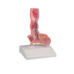
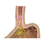
This life-size model shows a frontal section of the lower part of the oesophagus and the upper part of the stomach.The most common diseases are depicted.
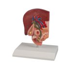
This half natural size model shows the anatomy of the biliary system and ist surroundings in great detail.
3B Scientific Smart Anatomy Gallstone Model shows the anatomy of the biliary system and its surroundings in half natural size.
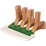
Four-piece model of progressive stages of GERD. Include: normal, sliding hiatal hernia and acid reflux; chronic acid reflux; Barrett’s esophagus/adeno?carcinoma.
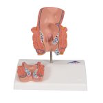
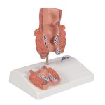
3B Scientific Smart Anatomy Hemorrhoid Model is a life-size frontal section of the rectum as well as a smaller relief on a pedestal.
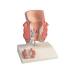
This descriptive model for patient education in about twice life size shows a frontal section through the rectum.
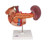
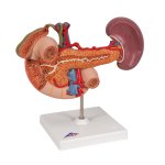
3B Scientific Smart Anatomy life-size Model of Rear Organs of Upper Abdomen shows the duodenum, gall bladder and bile ducts, the pancreas, the spleen and vessels.
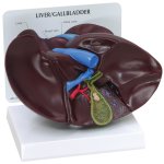
The liver-gallbladder model is a full size liver and gallbladder with cut-away section showing inner anatomy of gallbladder, including gallstones.
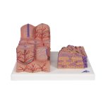
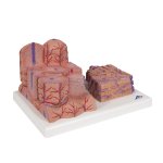
3B Scientific Smart Anatomy 3B MICROanatomy Liver Model shows a highly magnified diagrammatic view of a section of the liver.
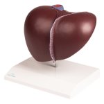
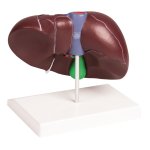
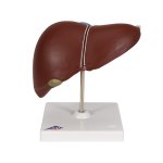
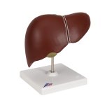
This realistic model reproduces a liver with the gall bladder. The hilus vessels are shown as well as the extrahepatic ducts and the main ligaments.
3B Scientific Smart Anatomy Liver Model with Gall Bladder shows: 4 lobes with gall bladder, Extrahepatic, ducts Hilus, vessels
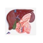
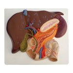
3B Scientific Smart Anatomy Liver Model with Gall Bladder, Pancreas & Duodenum. The anatomy of the liver with Gall Bladder, Pancreas and Duodenum is graphically illustrated.
This life size model shows a section of the liver with gall bladder, pancreas and duodenum; includes hepatic and pancreatic ducts.
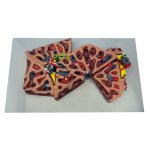
This greatly enlarged model shows the fine detail of a single liver lobule, which is sectioned and shown in relationship to portions of surrounding lobules.
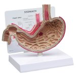
The Stomach Model with Ulcers is a full size cut-away section of stomach shows gastric ulcer, duodenal ulcer and esophageal inflammation.
Items 1 - 20 of 44


