

Musculature
Musculature models
GALAXYMED provides the most helpful tools in medical education. Anatomical models of the human musculature: Skulls with facial muscles, detailed muscles models e.g. of legs, arms, hands, feet, hips, knees, backs etc. The amazing life-size muscular figure with 39 parts of detachable muscles is with its accurate detail in high quality manufactured especially suitable for the high demands of medical schools.
Items 1 - 20 of 35

The base model "Foot with 2 exposed tendons, Achilles tendon, tibialis posterior tendon" from MEDICAL FX Germany is a handcrafted silicone model of a human foot featuring an exposed Achilles tendon (Tendo Achillis) and tibialis posterior tendon (Tendo musculi tibialis posterioris).

The "Base Model Arm with Exposed Tendons” from MEDICAL FX Germany is an anatomical representation specially designed to visualize the complex tendon structures of the human arm.


This life size model of a right human foot is medially opened and shows bones, joints, ligaments, fatty tissue and muscles.
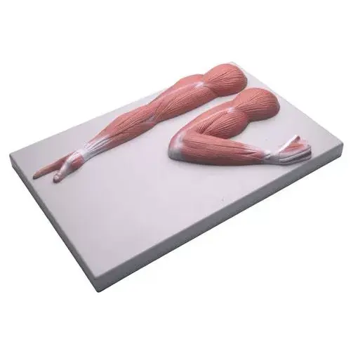
This model illustrates the muscles of the arm in the relaxed and contracted states. Muscles and tendons are showed in great detail. Mounted on base.


An invaluable tool for physicians who wish to discuss back pain with their patients, this back muscle model is designed for interaction and education.


This life size model is a ventral view of the human neck, showing excellent details of the main muscles, vascular structures, nerves and glands.

Full size normal muscled right knee model includes: rectus femoris, vastus lateralis and vastus medialis muscles; femur, fibula, patella and tibia bones; ligaments and tendons.

Full size right muscled hip model includes: gluteus medius, gluteus minimus, iliacus, inferior and superior gemellus, obturator internus, piriformis and psoas muscles.

Full size normal right elbow includes: biceps brachii, brachialis, pronator teres and supinator muscles; bones, ligaments and nerves

Full size normal right hip with proximal femur and lower lumbar vertebrae including: sciatic nerve, gluteus medius, gluteus minimus, iliacus, inferior and more


3B Scientific Smart Anatomy 3B MICROanatomy human muscle fiber model, 10,000 times magnified illustrates a section of a muscle fiber and its neuromuscular end plate.


This model has extraordinary fine structures and a very detailed painting. On one side it shows the superficial musculature, on the other side the deep muscles.


This model shows the muscles of the head, neck and superior thoracic region. Superfi cial and deep musculature are well represented, as are the pathways of the subclavian and carotid arteries.


This life size model shows the musculature oft he human arm in detail. It shows superficial and deep muscle structures, vessels, nerves and ligaments.


3B Scientific Smart Anatomy half head model with neck, muscles, blodd vessels & nerve branches represents the outer, superficial and the internal structures.
Top rated


3B Scientific Smart Anatomy Muscle Arm Model, 3/4 life-size, 6 part illustrates both the superficial and deeper muscles, five of which are removable from the muscled arm.


This life size model shows the musculature oft he human leg in detail. It shows superficial and deep muscle structures, vessels, nerves and ligaments.


3B Scientific Smart Anatomy 1/3 life-size human muscle figure, 2 part. All the superficial musculature is accurately reproduced and detailed in life like colors.


This model shows the muscles of the head, neck and superior thoracic region. Superfi cial and deep musculature are well represented, as are the pathways of the subclavian and carotid arteries.
Top rated


3B Scientific Smart Anatomy Muscle Leg Model, 3/4 life-size, 9 part. The muscular leg model illustrates both the superficial and deeper muscles, eight of which are removable.
Items 1 - 20 of 35