

Skin
Skin Models
Items 1 - 20 of 20

The Tablet Wound Model "4 Scar Model Keloid Scar, Atrophic Scar, Normal Scar, Hypertrophic Scar" by MEDICAL FX Germany combines four clinically relevant scar types on a single training model: the Normal Scar, the Atrophic Scar, the Hypertrophic Scar, and the Keloid Scar.

The wound model "Stick-On Medium Skin Surface Without Wounds” from MEDICAL FX Germany offers a unique opportunity to integrate intact human skin into simulations.

With this model it is easy to compare the structures of hairy and hairless skin: sensitive corpuscles, blood vessels, sweat gland, nerves, hair and hair root.


3B Scientific Smart Anatomy Human Skin Section Model, 70 times life-size. This unique skin model shows a section of human skin in three dimensional form.


3B Scientific Smart Anatomy Human Skin Section Model, 70 times life-size This skin relief model shows a section through the three layers of the hair-covered skin of the head. The skin section shows: Representation of hair follicles with sebaceous glands Sweat glands Receptors Nerves Vessels The skin section portrays the anatomy of the skin at 70 times life size. The skin model is delivered on base.


3B Scientific Smart Anatomy Human Skin Section Model, 40 times life-size features detail with hair follicles, sebaceous glands, sweat glands, receptor, nerves and vessels.


3B Scientific Smart Anatomy Human Skin Model, 80 times magnified, 3 part represents the three most important sections of skin: the human scalp, the palm and the back of a hand.


3B Scientific Smart Anatomy Skin Cancer Model with 5 stages, 8 times magnified shows healthy skin and 5 different stages of malignant melanoma on the front and back.


Model presents full size and enlarged views of dysplastic nevus, actinic keratosis, malignant melanoma, squamous cell carcinoma, and basal cell carcinoma skin lesions.


Enlarged two-sided skin cross-section. The burn side shows indication of first, second and third degree burns. The reverse side illustrates normal skin anatomy

This model of a human skin in about 50 times life size shows 3 dimensional the different skin layers and anatomical structures.


"Enlarged two-sided skin cross-section model. The ""acne"" side displays: whitehead, blackhead and pustule/cyst. The reverse side illustrates normal skin anatomy."

The Skin Acne model is an enlarged cylindrical cross-section of skin with relief detail displaying: acne pustule, closed comedo, and open comedo on backside.
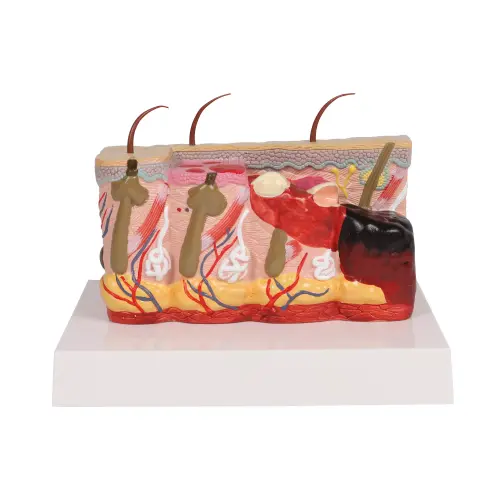

This model, approx. 30 times enlarged, compares normal skin with first, second and third degree burnt skin, showing also the subsequent pathological changes. The first level of injury shows epidermis damage and erythema; the second one includes hypodermis damage and vescicles, the third one reveals a subcutaneous damage.
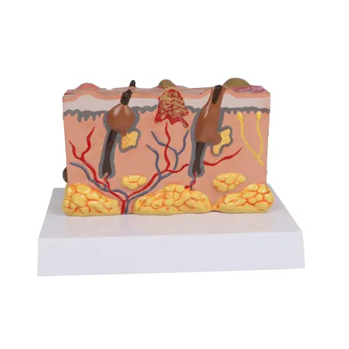

This model, approximately 30X life size, features, on the one side, detailed and realistic depictions of different closed and open lesions. On the other side the model shows cyst, fissure, pustule, ulcer, scale and blister. All the pathologies are showed separately in the model with a realistic representation of their morphology

This approximately 5 times enlarged model of the terminal part of a typical digit shows three sectional views of the nail root and bed, germinative region and bone.


3B Scientific Smart Anatomy Microscopic Skin, Hair & Nail Model shows the microscopic structure of the human skin in great detail.

The Skin Cancer Trainer has been developed to assist with educating health care providers about the various types of skin cancer and what they may look like.

The Human Skin Series with Burn Pathologies is enlarged 75 times life size to detail skin burn pathologies. Six skin models in one.


The „Frame Model Hidradenitis Suppurativa (Curved Cross-Section)” from MEDICAL FX Germany illustrates the typical progression of the chronic inflammatory skin disease hidradenitis suppurativa
Items 1 - 20 of 20