

Sensory Organs
Sensory Organs
Anatomic models of the sensory organs as eyes, ears and skin are widely used in teaching of Human Anatomy in universities, schools and colleges. They are also of use for patient education.


Items 1 - 30 of 30
Top rated


3B Scientific Smart Anatomy Human Eye Model, 3 times life-size, 7 part shows the optic nerve in its natural position in the bony orbit of the eye (floor and medial wall).


This 13-part model, magnified approximately 2.5x, shows the anatomy of the human eye. The eye socket can be divided into three parts and then shows the internal structures.


Model can be divided horizontally to show internal details. Cornea, iris, lens and vitreous body can be removed.


3B Scientific Smart Anatomy Human Eye Model, 3 times life-size, 6 part. Great model to teach-learn the anatomy of the human eye! Human eye on base.


Complex hand painted reproduction of a human eye in about 4 times life size.


3B Scientific Smart Anatomy Human Eye Model, 5 times life-size, 6 part. The Giant Eye replica is a great tool to teach-learn the anatomy of the eye!
Top rated


3B Scientific Smart Anatomy Human Eye Model, 5 times life-size, 7 part. Great for studying the anatomy of the human eye! Eye on base of bony orbit.


3B Scientific Smart Anatomy Organ of Corti Model with Representation in Cochlea shows a three dimensional section through the organ of Corti, the site of the sense of hearing.


Full size model of human ear is clear to aid viewing of ear canal, tympanic membrane, stapes, incus, malleus and cochlea of the inner ear.


3B Scientific The human auditory ossicles, individually presented in natural position, embedded in transparent acrylic. Cast from natural specimen.

This model of a human skin in about 50 times life size shows 3 dimensional the different skin layers and anatomical structures.

With this model it is easy to compare the structures of hairy and hairless skin: sensitive corpuscles, blood vessels, sweat gland, nerves, hair and hair root.


3B Scientific Smart Anatomy Human Skin Section Model, 70 times life-size. This unique skin model shows a section of human skin in three dimensional form.

The Hypertension Set includes a miniature brain, eye, heart, kidney and artery models. Education card illustrates effects associated with hypertension


Enlarged approx. 15 times. The model shows instructive the organs of the middle and inner ear. The bony and membranous labyrinths are shown and the cochlea can be opened.

This approximately 5 times enlarged model of the terminal part of a typical digit shows three sectional views of the nail root and bed, germinative region and bone.


3B Scientific Smart Anatomy Human Nose Model with Paranasal Sinuses, 5 part illustrates the structure in the upper right half of a face in 1.5-fold enlargement.

Full size normal model shows semi-circular canals and cochlea of the inner ear, auditory ossicles, tympanic membrane, temporal and tensor tympani muscles.


Child’s ear model illustrates: semi-circular canals and cochlea of the inner ear; auditory ossicles, otitis media; tympanic membrane, temporal and tensory tympani muscles


3B Scientific Smart Anatomy Human Ear Model for Desktop, 1.5 times life-size represents the outer, middle, and inner ear. Ear on base.

This slightly enlarged model of a human ear with all anatomical details shows the auditory canal, the tymphanic membrane, malleus, incus, stapes and cochlea.
Top rated


3B Scientific Smart Anatomy Human Ear Model, 3 times life-size, 4 part This high quality model of the human ear represents outer, middle and inner ear.


This model shows the details of the external, middle and inner ear in three times life size.


3B Scientific Smart Anatomy Human Ear Model, 3 times life-size, 6 part This high quality human ear model represents outer, middle and inner ear.

Ossicles are shipped embedded in a custom made foam insert inside a clear plastic petri dish. Ossicles can be removed easily.


3B Scientific Smart Anatomy Pathological Human Eye Model shows an eye enlarged by a factor of 5. Perfect for educating patients in the anatomy and changes of the human eye.
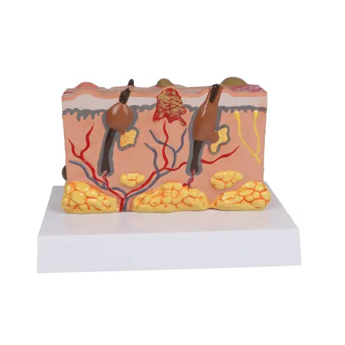

This model, approximately 30X life size, features, on the one side, detailed and realistic depictions of different closed and open lesions. On the other side the model shows cyst, fissure, pustule, ulcer, scale and blister. All the pathologies are showed separately in the model with a realistic representation of their morphology


A unique model specifically designed to aid understanding of the five special senses: sight, smell, taste, hearing, balance, with their related nerves.

Transparent 3-D construction makes it easy to understand the structure from auricle to drum.


This item shows the anatomy of the life-size tongue, including the palatin and lingual tonsils, epiglottis and muscles. The model also includes a detailed 20X life-size enlargement of the tongue tissue to show its histology: taste buds, lingual and mucous glands, crypt and muscles are well represented.
Items 1 - 30 of 30